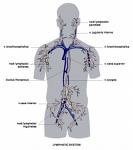
Lymphatic System
The lymphatic system in vertebrates is a network of conduits that carry a clear fluid called lymph. It also includes the lymphoid tissue through which the lymph travels. Lymphoid tissue is found in many organs, particularly the lymph nodes, and in the lymphoid follicles associated with the digestive system such as the tonsils. The system also includes all the structures dedicated to the circulation and production of lymphocytes, which includes the spleen, thymus, bone marrow and the lymphoid tissue associated with the digestive system.
The blood does not directly come in contact with the parenchymal cells and tissues in the body, but constituents of the blood first exit the microvascular exchange blood vessels to become interstitial fluid, which comes into contact with the parenchymal cells of the body. Lymph is the fluid that is formed when interstitial fluid enters the initial lymphatic vessels of the lymphatic system. The lymph is then moved along the lymphatic vessel network by either intrinsic contractions of the lymphatic vessels or by extrinsic compression of the lymphatic vessels via external tissue forces (e.g. the contractions of skeletal muscles.
The lymphatic system has three interrelated functions. It is responsible for the removal of interstitial fluid from tissues. It absorbs and transports fatty acids and fats as chyle to the circulatory system. The last function of the lymphatic system is the transport of immune cells to and from the lymph nodes. The lymph transports antigen presenting cells (APCs), such as dendritic cells, to the lymph nodes where an immune response is stimulated. The lymph also carries lymphocytes from the efferent lymphatics exiting the lymph nodes.
The study of lymphatic drainage of various organs is important in diagnosis, prognosis, and treatment of cancer. The lymphatic system, because of its physical proximity to many tissues of the body, is responsible for carrying cancerous cells between the various parts of the body in a process called metastasis. The intervening lymph nodes can trap the cancer cells. If they are not successful in destroying the cancer cells the nodes may become sites of secondary tumors.
Diseases and other problems of the lymphatic system can cause swelling and other symptoms. Problems with the system can impair the body's ability to fight infections.
Organization[]
The lymphatic system can be broadly divided into the conducting system and the lymphoid tissue.
The conducting system carries the lymph and consists of tubular vessels that include the lymph capillaries, the lymph vessels, and the right and thoracic ducts.
The lymphoid tissue is primarily involved in immune responses and consists of lymphocytes and other white blood cells enmeshed in connective tissue through which the lymph passes. Regions of the lymphoid tissue that are densely packed with lymphocytes are known as lymphoid follicles. Lymphoid tissue can either be structurally well organized as lymph nodes or may consist of loosely organized lymphoid follicles known as the mucosa-associated lymphoid tissue.
Formation of lymph[]
Formation of interstitial fluid from blood
Blood supplies nutrients and important metabolites to the tissues, and collects back the waste products that they produce, which requires exchange of respective constituents between the blood and tissues. This exchange is not direct, however, and is effected through an intermediary called interstitial fluid or tissue fluid that the blood forms. Interstitial fluid (ISF) is the fluid that occupies the spaces between the cells and acts as their immediate environment. As the blood and the surrounding cells continually add and remove substances from the ISF, its composition keeps on changing. Water and solutes can freely pass (diffuse) between the ISF and blood, and thus both are in dynamic equilibrium with each other; exchange between the two fluids occurs across the walls of small blood vessels called capillaries.
ISF forms at the arterial (coming from the heart) end of the capillaries because of higher pressure of blood, and most of it returns to its venous ends and venules; the rest (10—20%) enters the lymph capillaries as lymph.[1] Thus, lymph when formed is a watery clear liquid with the same composition as the ISF. As it flows through the lymph nodes, however, it comes in contact with blood and tends to accumulate more cells (particularly lymphocytes) and proteins.
The two primary lymph systems are the thymus gland and the bone marrow, where the immune cells form or mature. The secondary lymph system is made up of encapsulated and unencapsulated diffuse lymphoid tissue. The encapsulated tissue includes the spleen and the lymph nodes. The unencapsulated tissue includes the gut-associated lymphoid tissues and the tonsils.
Lymphoid tissue[]
Lymphoid tissue associated with the lymphatic system is concerned with immune functions in defending the body against the infections and spread of tumors. It consists of connective tissue with various types of white blood cells enmeshed in it, most numerous being the lymphocytes.
The lymphoid tissue may be primary, secondary, or tertiary depending upon the stage of lymphocyte development and maturation it is involved in. Primary (central) lymphoid tissues serve to generate mature virgin lymphocytes from immature progenitor cells. Secondary (peripheral) lymphoid tissues provide a place where lymphocytes can talk to each other; an environment for antigen focusing, where lymphocytes can 'study' an antigen and sharpen up the immune response by clonal expansion and affinity maturation; and provide a home for lymphocytes, where they can be available when they are needed.
The thymus and the bone marrow constitute the primary lymphoid tissues involved in the production and early selection of lymphocytes. Secondary lymphoid tissue provides the environment for the foreign or altered native molecules (antigens) to interact with the lymphocytes. It is exemplified by the lymph nodes, and the lymphoid follicles in tonsils, Peyer's patches, spleen, adenoids, skin, etc. that are associated with the mucosa-associated lymphoid tissue (MALT). The tertiary lymphoid tissue typically contains much fewer lymphocytes, and assumes an immune role only when challenged with antigens that result in inflammation. It achieves this by importing the lymphocytes from blood and lymph.
Lymph nodes[]
A lymph node is an organized collection of lymphoid tissue, through which the lymph passes on its way to returning to the blood. Lymph nodes are located at intervals along the lymphatic system. Several afferent lymph vessels bring in lymph, which percolates through the substance of the lymph node, and is drained out by an efferent lymph vessel.
The substance of a lymph node consists of lymphoid follicles in the outer portion called the "cortex", which contains the lymphoid follicles, and an inner portion called "medulla", which is surrounded by the cortex on all sides except for a portion known as the "hilum". The hilum presents as a depression on the surface of the lymph node, which makes the otherwise spherical or ovoid lymph node bean-shaped. The efferent lymph vessel directly emerges from the lymph node here. The arteries and veins supplying the lymph node with blood enter and exit through the hilum.
Lymph follicles are a dense collection of lymphocytes, the number, size and configuration of which change in accordance with the functional state of the lymph node. For example, the follicles expand significantly upon encountering a foreign antigen. The selection of B cells occurs in the germinal center of the lymph nodes.
Lymph nodes are particularly numerous in the mediastinum in the chest, neck, pelvis, axilla (armpit), inguinal (groin) region, and in association with the blood vessels of the intestines.
Lymphatics[]
lymphatic system
Tubular vessels transport back lymph to the blood ultimately replacing the volume lost from the blood during the formation of the interstitial fluid. These channels are the lymphatic channels or simply called lymphatics.[2]
General structure of Lymphatics[]
The general structure of lymphatics is based on that of blood vessels. There is an inner lining of single flattened cells composed of a type of epithelium that is called endothelium, and the cells are called endothelial cells. This layer functions to mechanically transport fluid and since the basement membrane on which it rests is discontinuous; it leaks easily. The next layer is that of smooth muscles that are arranged in a circular fashion around the endothelium, which by shortening (contracting) or relaxing alter the diameter (caliber) of the lumen. The outermost layer is the adventitia that consists of fibrous tissue. The general structure described here is seen only in larger lymphatics; smaller lymphatics have fewer layers. The smallest vessels (lymphatic or lymph capillaries) lack both the muscular layer and the outer adventitia. As they proceed forward and in their course are joined by other capillaries, they grow larger and first take on an adventitia, and then smooth muscles.
Unlike the cardiovascular system, the lymphatic system is not closed and has no central pump. Lymph movement occurs despite low pressure due to peristalsis (propulsion of the lymph due to alternate contraction and relaxation of smooth muscle), valves, and compression during contraction of adjacent skeletal muscle and arterial pulsation.
Lymph capillaries[]
The lymphatic circulation begins with blind ending (closed at one end) highly permeable superficial lymph capillaries, formed by endothelial cells with button-like junctions between them that allow fluid to pass through them when the interstitial pressure is sufficiently high.[3] These button-like junctions consist of protein filaments like platelet endothelial cell adhesion molecule-1 or (PECAM-1). A valve system in place here prevents the absorbed lymph from leaking back into the ISF. There is another system of semilunar (semi=half; lunar=related to the Moon) valves that prevents back-flow of lymph along the lumen of the vessel.[3] Lymph capillaries have many interconnections (anastomoses) between them and form a very fine network.[4]
Rhythmic contraction of the vessel walls through movements may also help draw fluid into the smallest lymphatic vessels, capillaries. If tissue fluid builds up the tissue will swell; this is called edema. As the circular path through the body's system continues, the fluid is then transported to progressively larger lymphatic vessels culminating in the right lymphatic duct (for lymph from the right upper body) and the thoracic duct (for the rest of the body); both ducts drain into the circulatory system at the right and left subclavian veins. The system collaborates with white blood cells in lymph nodes to protect the body from being infected by cancer cells, fungi, viruses or bacteria. This is known as a secondary circulatory system.
Lymph vessels[]
The lymph capillaries drain the lymph to larger contractile lymphatics, which have valves as well as smooth muscle walls. These are called the collecting lymphatics.[5] As the collecting lymph vessel accumulates lymph from more and more lymph capillaries in its course, it becomes larger and is called the afferent lymph vessel as it enters a lymph node. Here the lymph percolates through the lymph node tissue and is removed by the efferent lymph vessel. An efferent lymph vessel may directly drain into one of the (right or thoracic) lymph ducts, or may empty into another lymph node as its afferent lymph vessel. Both the lymph ducts return the lymph to the blood stream by emptying into the subclavian veins
The functional unit of a lymph vessel is known as a lymphangion, which is the segment between two valves. Since it is contractile, depending upon the ratio of its length to its radius, it can act either like a contractile chamber propelling the fluid ahead, or as a resistance vessel tending to stop the lymph in its place.
Function of the fatty acid transport system[]
Lymph vessels called lacteals are present in the lining of the gastrointestinal tract, predominantly in the small intestine. While most other nutrients absorbed by the small intestine are passed on to the portal venous system to drain, via the portal vein, into the liver for processing, fats (lipids) are passed on to the lymphatic system, to be transported to the blood circulation via the thoracic duct. The enriched lymph originating in the lymphatics of the small intestine is called chyle. As the blood circulates, fluid leaks out into the body tissues. This fluid is important because it carries food to the cells and waste back to the bloodstream. The nutrients that are released to the circulatory system are processed by the liver, having passed through the systemic circulation. The lymph system is a one-way system, transporting interstitial fluid back to blood.
Diseases of the lymphatic system[]
}}</ref> which may occur if the lymphatic system is damaged or has malformations. It usually affects the limbs, though face, neck and abdomen may also be affected. An estimated 170 million people develop lymphedema, which progresses in three stages:
Stage 1: Pressing the swollen limb leaves a pit that takes a while to fill back in. Because there is little fibrosis (hardening) it is often reversible. Elevation reduces swelling.
Stage 2: Pressure does not leave a pit. Elevation does not help. If left untreated, the limb becomes fibrotic.
Stage 3: This stage of lymphedema is often called elephantiasis. It is generally only in the legs after lymphedema that has gone long untreated. While treatment can help a little, it is not reversible.
Some common causes of swollen lymph nodes include infections, infectious mononucleosis, and cancer, e.g. Hodgkin's and non-Hodgkin's lymphoma, and metastasis of cancerous cells via the lymphatic system. In elephantiasis, infection of the lymphatic vessels cause a thickening of the skin and enlargement of underlying tissues, especially in the legs and genitals. It is most commonly caused by a parasitic disease known as lymphatic filariasis. Lymphangiosarcoma is a malignant soft tissue tumor (soft tissue sarcoma), whereas lymphangioma is a benign tumor occurring frequently in association with Turner syndrome. Lymphangioleiomyomatosis is a benign tumor of the smooth muscles of the lymphatics that occurs in the lungs.
Development of lymphatic tissue[]
Lymphatic tissues begin to develop by the end of the fifth week of embryonic life. Lymphatic vessels develop from lymph sacs that arise from developing veins, which are derived from mesoderm.
The first lymph sacs to appear are the paired jugular lymph sacs at the junction of the internal jugular and subclavian veins. From the jugular lymph sacs, lymphatic capillary plexuses spread to the thorax, upper limbs, neck and head. Some of the plexuses enlarge and form lymphatic vessels in their respective regions. Each jugular lymph sac retains at least one connection with its jugular vein, the left one developing into the superior portion of the thoracic duct.
The next lymph sac to appear is the unpaired retroperitoneal lymph sac at the root of the mesentery of the intestine. It develops from the primitive vena cava and mesonephric veins. Capillary plexuses and lymphatic vessels spread form the retroperitoneal lymph sac to the abdominal viscera and diaphragm. The sac establishes connections with the cisterna chyli but loses its connections with neighboring veins.
The last of the lymph sacs, the paired posterior lymph sacs, develop from the iliac veins. The posterior lymph sacs produce capillary plexuses and lymphatic vessels of the abdominal wall, pelvic region, and lower limbs. The posterior lymph sacs join the cisterna chyli and lose their connections with adjacent veins.
With the exception of the anterior part of the sac from which the cisterna chyli develops, all lymph sacs become invaded by mesenchymal cells and are converted into groups of lymph nodes.
The spleen develops from mesenchymal cells between layers of the dorsal mesentery of the stomach. The thymus arises as an outgrowth of the third pharyngeal pouch.
Video[]
thumb|300px|right