About[]
Diagnostic Ultrasound
By: Nate Curtis, Bio-Medical Electronics Student, Western Technical College, La Crosse, WI, 10-16-08
Diagnostic ultrasounds are invaluable pieces of medical equipment used by medical professionals on a daily basis. Most people think of diagnostic ultrasound as only being used to see a developing baby, so I will give many other uses of diagnostic ultrasound. In this article I will discuss what diagnostic ultrasound is, what basic parts are in a diagnostic ultrasound machine, and finally I will discuss different types of diagnostic ultrasound.
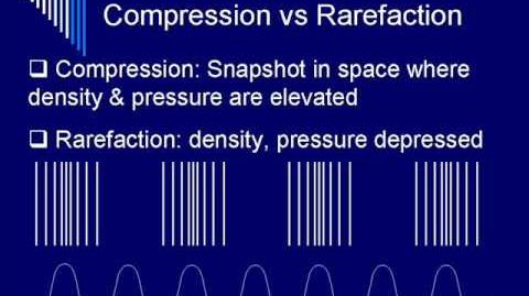
Diagnostic ultrasounds or ultrasonography is a medical imaging technique that uses high frequency sound waves and their echoes. The technique is very similar to the echolocation used by bats, whales and dolphins, as well as SONAR used by submarines. In diagnostic ultrasounds six actions need to happen. First, the ultrasound machine transmits high-frequency (1 to 20 megahertz) sound pulses into your body using a probe. Second, the sound waves travel into your body and hit a boundary between tissues (e.g. between fluid and soft tissue, soft tissue and bone). Next, some of the sound waves get reflected back to the probe, while some travel on further until they reach another boundary and get reflected. Then, the reflected waves are picked up by the probe and relayed to the ultrasound machine. Next, the ultrasound machine calculates the distance from the probe to the tissue or organ (boundaries) using the speed of sound in tissue (5,005 ft/s or 1,540 m/s) and the time of the each echo’s return, 13 microseconds per centimeter, round trip. Finally, the ultrasound machine displays the distances and intensities of the echoes on the screen, forming a two dimensional or three dimensional image.[1]
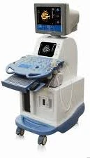
A typical diagnostic ultrasound setup has seven basic components. The first component is a ultrasound transducer probe. The transducer probe is the main part of the ultrasound machine. The transducer probe makes the sound waves and receives the echoes. The transducer probe generates and receives sound waves using a principle called the piezoelectric (pressure electricity) effect, which was discovered by Pierre and Jacques Curie in 1880. In the probe, there are multiple quartz crystals called piezoelectric crystals. When an electric current is applied to these crystals, they change shape rapidly. The rapid shape changes, or vibrations, of the crystals produce sound waves that travel outward. Conversely, when sound or pressure waves hit the crystals, they emit electrical currents. Therefore, the same crystals can be used to send and receive sound waves. The probe also has a sound absorbing substance to eliminate back reflections from the probe itself, and an acoustic lens to help focus the emitted sound waves. The second component in a basic diagnostic ultrasound setup is a central processing unit (CPU). The CPU is the brain of the ultrasound machine. The CPU is basically a computer that contains the microprocessor, memory, amplifiers and power supplies for the microprocessor and transducer probe. The CPU sends electrical currents to the transducer probe to emit sound waves, and also receives the electrical pulses from the probes that were created from the returning echoes. The CPU does all of the calculations involved in processing the data. Once the raw data are processed, the CPU forms the image on the monitor. The CPU can also store the processed data and/or image on disk. The third component is transducer pulse controls. The transducer pulse controls allow the operator to set and change the frequency and duration of the ultrasound pulses, as well as the scan mode of the machine. The commands from the operator are translated into changing electric currents that are applied to the piezoelectric crystals in the transducer probe. The fourth component of a diagnostic ultrasound setup is the display. The display is a computer monitor which shows the processed data from the CPU. Displays can be black-and-white or color, depending upon the model of the ultrasound machine. The fifth component is a keyboard/cursor. Diagnostic ultrasound machines have a keyboard and a cursor, such as a trackball, built in. These devices allow the operator to add notes to and take measurements from the data. The sixth component of a diagnostic ultrasound setup is disk storage. The processed data and/ or images can be stored on disk. The disks can be hard disks, floppy disks, compact discs or digital video discs. Typically, a patients ultrasound scans are stored on a disk and archived with the patients medical records. The seventh, and final, component is a printer. Many diagnostic ultrasound machines have thermal printers that can be used to capture a hard copy of the image from the display.[2]
Types[]
There are three main types of diagnostic ultrasounds; two- dimensional, 3D ultrasound imaging and Doppler ultrasound. Unlike with an x-ray, there is no ionizing radiation exposure with this test.[3]Two- dimensional ultrasound displays a two dimensional images, or slice, of a three dimensional object. 3D ultrasound imaging uses several two-dimensional images which are acquired by moving the probes across the body surface or rotating inserted probes. The two-dimensional scans are then combined by specialized computer software to form 3D images. 3D imaging allows you to get a better look at the organ being examined and is best used for early detection of cancerous and benign tumors, examining the prostate gland for early detection of tumors, looking for masses in the colon and rectum, and detecting breast lesions for possible biopsies. 3D imaging is also used for visualizing a fetus to asses its development, especially for observing abnormal development of the face and limb, and for visualizing blood flow in various organs or a fetus. Doppler ultrasound is based upon the Doppler Effect. When the object reflecting the ultrasound waves is moving, it changes the frequency of the echoes, creating a higher frequency if it is moving toward the probe and a lower frequency if it is moving away from the probe. How much the frequency is changed depends upon how fast the object is moving. Doppler ultrasound measures the change in frequency of the echoes to calculate how fast an object is moving. Doppler ultrasound has been used mostly to measure the rate of blood flow through the heart and major arteries.
Clinical Applications[]
Ultrasound has been used in a variety of clinical settings, including obstetrics and gynecology, cardiology and cancer detection. The main advantage of ultrasound is that certain structures can be observed without using radiation. Ultrasound can also be done much quicker than X-rays or other radiographic techniques. There are multiple uses of diagnostic ultrasound in obstetrics and gynecology. One use is to measure the size of the fetus to determine the due date. Some more uses are to determine the position of the fetus to see if it is in the normal head down position or breech, seeing the number of fetuses in the uterus, checking the sex of the baby, checking the fetus’s growth rate by making many measurements over time, and to determine whether there is an appropriate amount of amniotic fluid cushioning the baby. Finally, obstetrics and gynecology use diagnostic ultrasound to detect ectopic pregnancy, the life-threatening situation in which the baby is implanted in the mother’s Fallopian tubes instead of in the uterus. Cardiology uses diagnostic ultrasound to see the inside of the heart to identify abnormal structures or functions and to measure blood flow through the heart and major blood vessels. Urology also uses diagnostic ultrasound. Diagnostic ultrasound is used to measure blood flow through the kidney, to see kidney stones, and to detect prostate cancer early. In addition to these areas, there is a growing use for ultrasound as a rapid imaging tool for diagnosis in emergency rooms. Dr. Chris Moore, Yale Medical School has created several presentations on ultrasound for emergency care.[4]
In conclusion diagnostic ultrasound has been used for over 70 years. Karl Theo Dussik, a neurologist/ psychiatrist at the University of Vienna, Austria, who had begun experiments in the late 1930s, was generally regarded as the first physician to have employed ultrasound in medical diagnosis. There have been many concerns about the safety of ultrasound. Because ultrasound is energy, the question becomes “What is this doing to my tissues or my baby?” There have been some reports of low birth weight babies being born to mothers who had frequent ultrasound examinations during pregnancy. However, there have been no substantiated ill-effects of ultrasound documented in studies in either humans or animals. This being said, ultrasound should still be used only when necessary. I believe that the future of diagnostic ultrasound is limitless. I think that the computers will become faster and have more memory for storing data. I believe the entire ultrasound machine will become smaller, perhaps even hand-held for use in the field. I think that 3D ultrasounds will become sharper in image because of smaller and more insertable probes. I also believe that there will be new technology hitting the market such as GE’s 4D ultrasound. 4D ultrasound is similar to 3D ultrasound except 4D show animation or movement. The exciting thing with all this new advancement is that there is a lot of equipment that will need servicing and it will only add to my job security.[5]
Manufacturers[]
ATL (Now Philips)
General Electric (GE) (Also Vingmed, Medison and Voluson)
Philips (Also ATL and HP)
Acuson (Now Siemens)
Mindray
Sonoscape
Siemens
Biosound
Sonosite
Toshiba
Hitachi
Models[]
Vivid 7. Logiq 5, 7, 9. LogiqBook, Voluson 730. logig 400 - General Electric (GE)
Sequoia, Aspen, S2000 - Acuson (Siemens)
Sonos 5500, 7500 - HP (Philips)
iU22, iE33 - Philips
DC7 - Mindray
M5, M7 - Mindray
HDI 3000, HDI 5000. - ATL (Philips)
Aplio XV - Toshiba
Megas, Technos - Biosound
Second Source Parts[]
Second Source Service[]
Specialized Technologies
Conquest Imaging
References[]
- ↑ Freudenrich,Ph.D., Craig. "How Ultrasound Works." How Stuff Works. 10 Apr. 2008 <http://health.howstuffworks.com/ultrasound.htm>. 3. "Ultrasound." Wikipedia. 10 Apr. 2008 <http://en.wikipedia.org/wiki/Ultrasound>
- ↑ Woo, Dr. Joseph. "A Short History of the Development of Ultrasound in Obstetrics and Gynecology." Obstetric Ultrasound. 10 Apr. 2008 <http://www.ob-ultrasound.net/history1.html>
- ↑ FDA. Medical Imaging: Ultrasound. Page Last Updated: 06/06/2012. http://www.fda.gov/Radiation-EmittingProducts/RadiationEmittingProductsandProcedures/MedicalImaging/ucm115357.htm
- ↑ Introduction to Point-of-care Ultrasound (POCUS) <http://ultrasound.guide/course?courseid=5001>
- ↑ "4D View for Voluson." GE Healthcare. 2008. General Electric. 10 Apr. 2008 <http://www.gehealthcare.com/usen/ultrasound/genimg/products/voluson730/4dview.html>
Video[]
Physics of Diagnostic Ultrasound - Segment One
Physics of Diagnostic Ultrasound - Segment One

Physics of Diagnostic Ultrasound - Segment Two
Physics of Diagnostic Ultrasound - Segment Two